

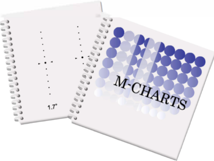
The M-Chart (Modified Cheltenham Auto-Steriogram / Membrane Differential Quantification Chart) is an excellent supplementary test for the quantitative and objective measurement of metamorphopsia (image distortion/warping) experienced by patients, particularly in cases of macular disease (e.g., epiretinal membrane, macular hole, age-related macular degeneration, and central serous chorioretinopathy).
While the Amsler Grid is effective at detecting the presence of metamorphopsia, it cannot numerically measure the degree of distortion. The M-Chart, however, allows you to clearly grade the patient's metamorphopsia score (M-score) in both vertical and horizontal planes.
The following steps can be taken to integrate the M-Chart into diagnostic processes in an eye clinic:
1. Triage and Administration Protocol
Which Patients Should Be Tested?
All patients presenting with complaints of metamorphopsia or central visual disturbance.
Patients with disrupted foveal anatomy on OCT (e.g., ERM, vitreomacular traction, CSCR, neovascular AMD).
Patients scheduled for or who have undergone macular surgery (e.g., ERM peeling or macular hole surgery).
How Is It Administered?
The test is administered monocularly at a distance of approximately 30 cm, with the patient wearing their near-vision correction (reading glasses).
The patient is asked to indicate when the dotted lines appear as a straight line.
Separate assessments are performed for both vertical and horizontal lines to obtain the vertical M-score ($M_v$) and the horizontal M-score ($M_h$).
2. Integration into Diagnostic and Follow-up Processes
Correlation and Cross-Validation with OCT:
Retinal Thickness vs. Functional Loss: Sometimes, despite a prominent ERM visible on OCT, the patient may experience minimal functional discomfort (or vice versa). The M-Chart helps correlate the anatomical finding with functional symptoms.
Indicator of Foveal Involvement: An M-score of 0.5 or higher generally indicates significant impact on the foveal structure and a decline in the patient's quality of daily life.
Surgical Timing and Indications:
Relying solely on visual acuity (Snellen/ETDRS) when deciding on surgery—particularly for Epiretinal Membrane (ERM) cases—can be misleading; a patient may experience severe metamorphopsia even with 0.8 visual acuity.
An elevated preoperative M-score provides concrete, objective data that supports the surgical indication and can be shared with the patient.
Postoperative and Post-treatment Follow-up:
3. Use in Patient Communication
Patients often struggle to describe the sensation of "distorted vision." The M-Chart significantly enhances patient compliance and satisfaction by allowing the patient to see the numerical score corresponding to their complaint and providing concrete evidence of the progress made after treatment.
