
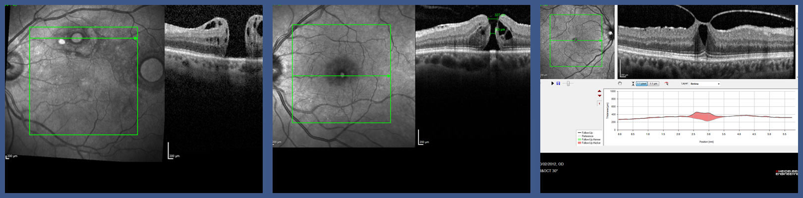
With OCT, each of the retina’s distinctive layers can be seen, allowing your ophthalmologist to map and measure their thickness. These measurements help with diagnosis and provide treatment guidance for glaucoma and retinal diseases, such as age-related macular degeneration and diabetic eye disease.
WHAT HAPPENS DURING OCT ?
You will be seated in front of the OCT machine and will rest your head on a support to keep it motionless. The equipment will then scan your eye without touching it. Scanning takes about 5 to 10 minutes. If your eyes were dilated, they may be sensitive to light for several hours after the exam.
WHAT CONDITIONS CAN OCT HELP TO DIAGNOSE ?
In addition, OCT is often used to evaluate disorders of the optic nerve. The optic nerve is made up of many nerve fibers and sends signals from your retina to your brain, where these signals are interpreted as the images you see. The OCT exam is helpful in determining changes to the fibers of the optic nerve, such as those caused by glaucoma.
Since OCT relies on light waves, it cannot be used successfully with any condition that interferes with light passing through the eye, such as dense cataracts or significant bleeding in the vitreous (the gel in the center of the eye).
