
 Ocular ultrasound, also known as ocular echography, “echo,” or a B-scan, is a quick, non-invasive test routinely used in clinical practice to assess the structural integrity and pathology of the eye. It can provide additional information not readily obtained by direct visualization of ocular tissues, and it is particularly useful in patients with pathology that prevents or obscures ophthalmoscopy, e.g. large corneal opacities, dense cataracts, or vitreous hemorrhage (1).
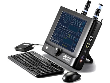
Ultrasound images can be obtained through the patient’s eyelids (as depicted in this tutorial) or with the probe directly on the surface of the eye with appropriate topical anesthesia.
The on-call ophthalmologist must be proficient at ocular ultrasound, as it is an indispensible tool for the diagnosis and triage of ophthalmic emergencies. One can systematically examine the entire globe with just five maneuvers, i.e. four dynamic quadrant views and one longitudinal cut through the macula and disc. One must always remember that this is simply a starting point, and a more detailed, comprehensive ultrasound examination should be guided by additional clinical data and preliminary ultrasonographic findings.
